www.fluorescencemicroscopy.it
Main menu:
- Home Page
- Microscopes
- Fluorescence
- Description
- Diascopy
- Epi-Fluoresc.
- Illumination
- Fluorocromes
- The Fading
- Wavelengths
- Properties
- % of transmission
- Types of filters
- Overlapping Spectra
- Intensity
- Image acquisition
- The Filter's set
- The Filters
- Fluorochrome's praparation
- Sample's preparations
- Fluorescence Sample
- Cytochemical Fluorescence
- Intrinsic Fluorescence
- Gallery
- Phase Contrast
- Polarization
- Darkfield
- DIC
- COL
- Rheinberg
- Brightfield
Intrinsic Fluorescence
Fluorescence
FLUORESCENCE APPLICATIONS IN SPECIMEN ANALYSIS :
INTRINSIC FLUORESCENCE
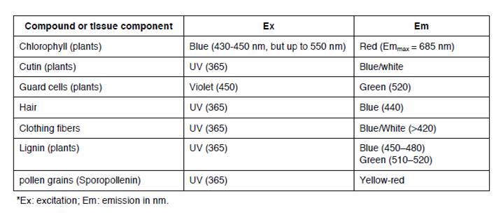
Most plant and some animal tissues exhibit intrinsic fluorescence (autofluorescence) attributed to the presence of secondary metabolites. Usually an obscuring phenomenon, intrinsic fluorescence may be used as a diagnostic tool for the identification of organ anatomy, cell constituents, or pathogenic activity.
Table 1. Intrinsic Fluorescence May Be Used as a Diagnostic Tool to Identify or Localize Certain Cell Components*
INDUCED BACKGROUND FLUORESCENCE
Aldehyde fixation, especially glutaraldehyde, induces a tissue fluorescence that spans the visible spectrum,
although it is especially bright in the yellow?red range. This induced fluorescence may obscure the localization of a fluorescent probe and may lead to false positive identification. When possible use a fluorescein-like probe
(fluorescein, Oregon Green, Cy2, BODIPY Fl, etc.) since its fluorescence emission may be distinguished from
background. Formaldehyde-induced-fluorescence (FIF) has been used as a diagnostic tool to distinguish certain
cell components and compounds. Treating tissue with either aqueous or heated formaldehyde vapors induces
autofluorescence that is often a different fluorescent color from endogenous and/or applied fluorescent probes.
INDUCED SPECIMEN FLUORESCENCE
Some dyes can be applied to tissue sections or whole mounts to induce a generalized fluorescence, which can
then be used in histochemical investigations. For example Eosin Y can be used on mammalian tissues to identify
fibers and other structures via fluorescence. Safranin O, Aniline Blue, the Acridine and fuchsin dyes, and myriad
other general purpose dyes can be used for fluorescence investigations.
Sub-Menu:
- Description
- Diascopy
- Epi-Fluoresc.
- Illumination
- Fluorocromes
- The Fading
- Wavelengths
- Properties
- % of transmission
- Types of filters
- Overlapping Spectra
- Intensity
- Image acquisition
- The Filter's set
- The Filters
- Fluorochrome's praparation
- Sample's preparations
- Fluorescence Sample
- Cytochemical Fluorescence
- Intrinsic Fluorescence ←
- Gallery